
Retrograde intra-renal surgery for stone extraction
RIRS is currently considered to be a safe standard retrograde endoscopic procedure for treating renal calculi. However, patients with stones>20 mm in diameter or multiple small calculi, especially in the presence of pre-existing tubes or following prior urinary tract infections, represent a subgroup of patients that are, in general, at higher risk of remarkable infectious complication rates and are likely to experience less satisfying stone-free rates when RIRS surgery is performed.
RIRS (retrograde intrarenal surgery):
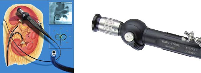
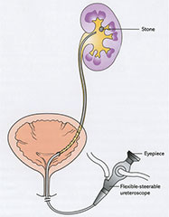
Retrograde intrarenal surgery (RIRS) is a procedure for doing surgery within the kidney using a viewing tube called a fiberoptic endoscope. In RIRS the scope is placed through the urethra (the urinary opening) into the bladder and then through the ureter into the urine-collecting part of the kidney. The scope thus is moved retrograde (up the urinary tract system) to within the kidney (intrarenal).
RIRS may be done to remove a stone. The stone is seen through the scope and can then be manipulated or crushed by an ultrasound probe or evaporated by a laser probe or grabbed by small forceps, etc.
RIRS is performed by a specialist, a urologist (endourologist) with special expertise in RIRS. The procedure is usually done under general or spinal anesthesia.
The advantages of RIRS over open surgery include a quicker solution of the problem, the elimination of prolonged pain after surgery, and much faster recovery.
Equipment & Techniques
The following list details the authors’ preferred equipment and materials for ureterorenoscopy.
- Fluoroscopy ( C-arm).
- Ureteroscopes, rigid/flexible.
- Guidewires (a variety).
- 5 fr ureteric catheter.
- Contrast medium.
- Ureteric access sheath.
- Holmium :YAG laser (with laser fibre, 365 or 200 ìm).
Flexible Ureteroscopy and Stone Fragmentation
This leaflet answers questions many people ask about ureteroscopy and stone fragmentation. If any further questions are raised from reading this information, please speak to members of the medical and/or nursing staff before, during or after your stay in hospital.
What does the procedure involve?
Telescopic removal/ fragmentation of stone(s) in the ureter or kidney with possible placement of a soft plastic tube or stent between the kidney and the bladder. This procedure also includes cystoscopy (looking inside the bladder with a telescope) and x-ray screening.
What are the alternatives to this procedure?
Open surgery, shock wave therapy or observation to allow spontaneous passage.
What should I expect before the procedure?
You will usually be admitted on the same day as your surgery. You will normally receive an appointment for pre-assessment, approximately 14 days before your admission, to assess your general fitness, to screen for the carriage of MRSA and to perform some baseline investigations. After admission, you will be seen by members of the medical team which may include the Consultant, Specialist Registrar, House Officer and your named nurse. An X-ray may be taken in advance of surgery to confirm the position of your stone(s).
You will be asked not to eat or drink for 6 hours before surgery and, immediately before the operation, you may be given a pre-medication by the anaesthetist which will make you dry-mouthed and pleasantly sleepy.
Please be sure to inform your Urologist in advance of your surgery if you have any of the following:
- An artificial heart valve
- A coronary artery stent
- A heart pacemaker or defibrillator
- An artificial joint
- An artificial blood vessel graft
- A neurosurgical shunt
- Any other implanted foreign body
- A prescription for Warfarin, Aspirin or Clopidogrel (Plavix®)
- A previous or current MRSA infection
What happens during the procedure?
Normally, a full general anaesthetic will be used and you will be asleep throughout the procedure.
You will usually be given injectable antibiotics before the procedure, after checking for any allergies.
A telescope is inserted into the bladder through the water pipe (urethra). Under X-ray screening, a flexible guidewire is inserted into the affected ureter up to the kidney. A longer telescope (either rigid or flexible) is then inserted into the ureter and passed up to the kidney. The stone is disintegrated using a mechanical probe or laser and the fragments extracted with special retrieval devices. If the surgeon feels it necessary a ureteric stent (tube to drain the kidney into the bladder) will be inserted.
What happens immediately after the procedure?
If a bladder catheter has been inserted, this is usually removed on the day after surgery. You will be able to go home once you are passing urine normally. An X-ray is often performed the day after surgery to check on the presence of residual stone fragments. The average hospital stay is 1 day.
Are there any side-effects?
Although the side-effects are listed below the majority of patients experience very mild symptoms which settle down spontaneously after a ureteroscopy.
Common
Mild burning or bleeding on passing urine for short period after operation Temporary insertion of a bladder catheter Insertion of a stent with a further procedure to remove it
If you have a ureteral stent, you may have:
- Blood in the urine: You may pass some blood clots in your urine right after the procedure. This usually stops after 3-4 times of passing urine. Blood in your urine may continue on and off until the stent is removed. The more active you are, the more blood you may see.
- The need to pass urine more often (frequency): The stent may give you the feeling that your bladder is full and you need to urinate. You may feel you have to urinate all the time, in small amounts. It will take your body a few days to adjust to the stent.
- Some abdominal/back discomfort (below the ribs) when you pass urine. This may decrease as your body adjusts to the stent. You may also feel bladder spasms when you pass urine.
- Nausea: The stent often causes a feeling of fullness. This makes some patients feel nauseated and sometimes vomit. This feeling will usually pass.
Occasional
- Inability to retrieve the stone or movement of the stone back into kidney where it is not retrievable.
- Kidney damage or infection needing further treatment.
- Failure to pass the telescope if the ureter is narrow.
- Recurrence of stones.
Rare
Damage to the ureter with need for open operation or tube placed into kidney directly from back (nephrostomy) to allow any leak to heal Very rarely, scarring or stricture of the ureter requiring further procedures.
What should I expect when I get home?
When you get home, you should drink twice as much fluid as you would normally to flush your system through and minimise any bleeding.
You may experience pain in the kidney over the first 24-72 hours, due to the swelling caused by insertion of the instrument or by the presence of a stent. Anti-inflammatory painkillers will help this pain which normally settles after 72 hours.
It will take at least 10 days to recover fully from the operation. You should not expect to return to work within 7 days.
What else should I look out for?
If you develop a fever, severe pain on passing urine, inability to pass urine or worsening bleeding, you should contact your GP immediately. Small blood clots or stone fragments may also pass down the ureter from the kidney, resulting in renal colic; in this event, you should contact your GP or attend the Accident and Emergency department.
Are there any other important points?
If a stent has been inserted, you will be informed before your discharge when the stent needs to be removed. Ureteric stents are usually removed under local anaesthetic. If you are unsure please check with your Urologist regarding the proposed date for the removal of the stent.
Stones have a tendency to recur so it you should ensure that you take adequate fluid (atleast 2.5 to 3 litres) everyday to reduce the chance of it recurring.
 WhatsApp Us